Mitochondrial Protein Import Advances the Case for Creation
What is true of economies is also true of cells. That is, any breakdown in the supply chain disrupts the efficient movement of cargo across boundaries. The impact of the pandemic on the US economy illustrates this reality.
One of the most enduring images of 2021 has to be the long line of cargo ships sitting off the Southern Californian coast for weeks on end, waiting to unload their containers at the Ports of Los Angeles and Long Beach.
These ports, which rank as America’s two busiest, handle about 40 percent of the country’s imports. Their economic importance can’t be underestimated. The US imports about $2.8 trillion dollars’ worth of goods each year.
This backlog wreaked havoc on the US economy during the summer and fall of 2021 and the reverberations are still being felt. The bottleneck in the supply chain made it difficult for American consumers to find many of the products we assume will always be readily available.
Economists identified at least two contributing factors to this shipping logjam.
- The COVID-19 pandemic led to a change in consumer purchasing habits. Instead of spending money on vacations, eating at restaurants, going to movies and shows, etc., American consumers began using their disposable income to purchase goods. This change in behavior dramatically increased the demand across the board for many goods and the supply chain simply wasn’t designed to handle a dramatic increase. Most of the products we buy are made overseas and must be shipped to the US.
- As the number of containers arriving at these two ports increased, there wasn’t enough capacity to process them. There were too few dock workers and not enough warehouse space to store the goods once unloaded. And there weren’t enough trains and trucks to transport the goods from the dock warehouses to their final destinations.
This breakdown in the supply chain illustrates how important the efficient movement of goods across borders is for many of the world’s economies.
The same is true for cells. To sustain many of the metabolic processes that drive cellular economies, the cell’s machinery must be able to move proteins and other types of cellular cargo across cell membranes—the cell’s boundaries.
This requirement is particularly true for protein import into mitochondria. If protein import into this organelle is disrupted, it will impair mitochondrial function and can result in serious diseases such as Parkinson’s and Alzheimer’s.
Researchers from the University of Freiburg in Germany have discovered that a protein dubbed DYRK1A regulates protein import into mitochondria by interacting with the TOM (translocase of the outer membrane) complex.1 The TOM complex consists of several distinct types of protein subunits and resides in the outer membrane of mitochondria. TOM is responsible for the initial steps of protein import into this organelle.
This insight helps explain previous observations that have shown that mutations to the gene that encodes DYRK1A correlate with the onset of Down’s syndrome, microcephaly, and autism. Mutations to the gene encoding DYRK1A will impair mitochondrial function, likely contributing to the etiology of these disorders.
This work also bears on evolutionary explanations for the origin of eukaryotic cells, specifically, the origin of mitochondria. The University of Freiburg team’s insight makes it even less likely that unguided evolutionary processes can account for the origin of eukaryotic cells, while at the same time making it much more credible to think that this key transition in life’s history resulted from a Creator’s handiwork.
Protein Import into Mitochondria
Save for a few exceptions, most mitochondrial proteins are made in the cell’s cytosol and then transported into mitochondria. The overall process of mitochondrial protein biogenesis consists of four stages: (1) protein synthesis, (2) targeting the protein to the mitochondria, (3) transporting the protein into the mitochondrial lumen, and (4) targeting the protein to its destination in the organelle.
The cell’s machinery initially makes mitochondrial proteins as pre-proteins with a signal sequence at one of its ends (the N terminus). The signal sequence has a specialized structure (an amphipathic α-helix) that serves to target the proteins to mitochondria. Think of the signal sequence as analogous to the shipping label on a container that lets the dock workers know which warehouse should receive the container. Receptor proteins that are part of the TOM complex recognize the signal sequence and transport the protein through a channel within the TOM interior into the intermembrane space (the region between the mitochondrial inner and outer membranes). Proteins called chaperones keep the mitochondrial proteins unfolded and stabilized throughout this process.
Figure: Protein Transport into Mitochondria
Credit: Heather Lanz
Once in the intermembrane space, another complex, TIM23, ushers the protein into the lumen (or the matrix) of the mitochondria. If the protein is to remain within the lumen (because that’s where it performs its work), then proteins called peptidases remove the signal sequence, and the protein adopts its intended three-dimensional shape.
If the protein is to be incorporated into the inner membrane, it possesses an additional targeting sequence that is recognized by two different protein complexes dubbed TIM22 and OXA. These biomolecular ensembles insert the protein into the inner membrane.
If the protein is to carry out its work in the intermembrane space, then the OXA complex will transport the protein back across the inner membrane. Alternatively, some proteins destined to operate in the inner membrane space possess a stop signal sequence. These sequences prevent the TIM23 complex from transporting the protein across the inner membrane into the lumen. Instead, peptidases in the intermembrane space remove the signal sequence, allowing the protein to adopt its operational structure.
Finally, if the protein is to be incorporated into the outer membrane, then another complex referred to as SAM (sorting and assembly machinery) inserts it into the outer membrane.
The Role of DYRK1A
Until recently, most biochemists viewed TOM as a passive pore with its channel always open. Biochemists now understand that the TOM channel can exist in both open and closed states depending on the cell’s metabolic state and its response to cellular “stress.”
As it turns out, the team from the University of Freiberg discovered that DYRK1A plays a role in controlling the entry of proteins into mitochondria by modifying TOM. DYRK1A is a (dual-specificity) protein kinase. These types of enzymes add phosphate groups to proteins. In this case, DYRK1A phosphorylates TOM70, one of the protein subunits that make up TOM. TOM70 is a protein receptor. As a protein receptor, it uses signal sequences to identify proteins that are slated for import into mitochondria and prepares them for their journey.
Phosphorylation of TOM70 makes TOM more effective at moving protein cargo into mitochondria. When TOM70 becomes de-phosphorylated, the rate of protein import into the mitochondria slows.
Even though TOM70 is part of TOM, it is loosely associated with TOM’s core (which is made up of the TOM40, TOM22, TOM5, TOM6 and TOM7 protein subunits). The phosphorylation state of TOM70 determines how strongly TOM70 interacts with the TOM core. When it is phosphorylated, TOM70 interacts strongly with the TOM core, facilitating protein transport. When de-phosphorylated, TOM70 interacts weakly with the TOM core, slowing down the movement of proteins into this organelle.
The University of Freiberg team learned that if DYRK1A is lost due to mutation or is inhibited, mitochondrial function becomes impaired. The researchers discovered that when either of these two scenarios takes place, the cell attempts to compensate by trying to increase the production of DYRK1A and the TOM proteins.
This biochemical response highlights the critical importance of regulating protein transport into mitochondria. These organelles are not only responsible for energy production in the cell, but they play a key role in the biosynthesis of key metabolites and cofactors, and they are also responsible for initiating programmed cell death (called apoptosis).
The discovery of this sophisticated protein transport system carries obvious biomedical significance. But it also has important implications for the leading evolutionary account for the origin of mitochondria, the endosymbiont hypothesis.
The Endosymbiont Hypothesis
(Note: Readers who are familiar with endosymbiogenesis should feel free to skip ahead to Scientific Challenges for Endosymbiogenesis.)
Russian botanist Konstantin Mereschkowsky initially proposed this model in the early 1900s and biologist Lynn Margulis advanced it in the late 1960s. Today, the endosymbiont hypothesis is widely assumed to be the explanation for the origin of eukaryotic cells. According to this idea, complex cells originated when symbiotic relationships formed among single-celled microbes after free-living bacterial and/or archaeal cells were engulfed by a “host” microbe.
Much of the work on the endosymbiont hypothesis centers around the origin of mitochondria. In some respects, the focus on the evolutionary origin of this organelle has become emblematic of the endosymbiont hypothesis. Presumably, the organelle started as an endosymbiont. Evolutionary biologists believe that once engulfed by the host cell, this microbe took up permanent residency, growing and dividing inside the host. Over time, the endosymbiont and the host became mutually interdependent, with the endosymbiont providing a metabolic benefit for the host cell, such as supplying a source of ATP. In turn, the host cell provided nutrients to the endosymbiont. Presumably, the endosymbiont gradually evolved into an organelle through a process referred to as genome reduction. This reduction resulted when genes from the endosymbiont’s genome were transferred into the genome of the host organism.
Evidence for the Endosymbiont Hypothesis
For evolutionary biologists, at least three lines of evidence bolster the endosymbiotic origin of mitochondria:
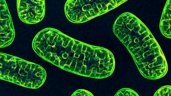
The similarity of mitochondria to bacteria. Most of the evidence for the endosymbiont hypothesis centers around the fact that mitochondria are about the same size and shape as a typical bacterium and have a double membrane structure like gram-negative cells. These organelles also divide in a way that is reminiscent of bacterial cells.
Mitochondrial DNA. Evolutionary biologists view the presence of the diminutive mitochondrial genome as a vestige of this organelle’s evolutionary history. They see the biochemical similarities between mitochondrial and bacterial genomes as further evidence for the evolutionary origin of these organelles.
The presence of the unique lipid, cardiolipin, in the mitochondrial inner membrane. This important lipid component of bacterial inner membranes is not found in the membranes of eukaryotic cells—except for the inner membranes of mitochondria. In fact, biochemists consider cardiolipin a signature lipid for mitochondria and another relic from its evolutionary past.
Scientific Challenges for Endosymbiogenesis
Despite the impressive collection of evidence for the evolutionary origin of mitochondria, endosymbiogenesis faces some significant scientific hurdles. (I have detailed some of these problems elsewhere. See the articles listed under Resources for Further Exploration.)
One of the most significant challenges involves explaining the origin of protein transport into the mitochondria.
The Protein Import Challenge to the Endosymbiont Hypothesis
Because we have some understanding as to why the import of goods into the US through the Ports of Los Angeles and Long Beach experienced a logjam, we can appreciate why an evolutionary origin of protein import into mitochondria is such a challenging problem. Each stage of mitochondrial protein biogenesis involves multiple steps, with each one carried out by an ensemble of proteins. Moreover, each step of the process must be precisely integrated with the other steps. If not, the entire process of mitochondrial protein biogenesis fails. To put it another way, each step involves an irreducibly complex biochemical apparatus which, in turn, integrates with the others to form the integrated complexity of the protein import pathway into mitochondria. That is, mitochondrial protein biogenesis can be characterized as an integrated, hierarchical, multilayered ensemble of irreducibly complex systems.
For mitochondrial protein biogenesis to emerge from an evolutionary standpoint, multiple biochemical systems had to originate simultaneously and become integrated with one another. For example, once mitochondrial genes became incorporated into the host genome, DNA sequences specifying signal sequences had to evolve and become precisely appended to every one of the mitochondrial DNA sequences. The TOM, TIM22, and TIM23 complexes had to evolve simultaneously to recognize mitochondrial proteins and work in tandem to move proteins into the mitochondria. In addition, chaperones had to emerge that would recognize mitochondrial proteins and keep them unfolded during the transport process. Signal peptidases had to evolve to remove signal sequences from mitochondrial proteins with exacting precision. Finally, stop sequences and additional targeting sequences had to evolve and become precisely positioned within the mitochondrial protein genes. And now, added to this list of requirements is the simultaneous origin of the DYRK1A protein kinase to regulate protein import in response to the cell’s metabolic status.
In other words, these demands are the same kinds of requirements that must simultaneously be met to successfully import goods into a country. If a system to unload containers, move the containers’ contents to a warehouse, and transport the warehouses’ contents to their destination isn’t all in place (or isn’t working efficiently), then the import process will collapse.
Evolutionary biologists have no inkling of how mitochondrial protein biogenesis could have evolved. According to cell biologist Franklin Harold, “The origin of the machinery for protein import is more complicated and is subject to much debate . . . Most of the transferred genes continue to support mitochondrial functions, having somehow acquired the targeting sequences that allow their protein products to be recognized by TOM and TIM and imported into the organelle.”2
To say that “the origin of the machinery for protein import” is a “complicated” system that “somehow” evolved is not a scientific explanation for how this complex biochemical system arose. It isn’t even an evolutionary just-so story. Instead, it is precisely what a creation model proponent would say based on what we have learned—and continue to learn—about protein import into mitochondria. The “somehow” factor that puzzles some scientists points to the work of an intelligent designer.
Resources for Further Exploration
Challenges to the Endosymbiont Hypothesis
“Evolutionary Paradigm Lacks Explanation for Origin of Mitochondria and Eukaryotic Cells” by Fazale Rana (article)
“Complex Protein Biogenesis Hints at Intelligent Design” by Fazale Rana (article)
“ATP Transport Challenges the Evolutionary Origin of Mitochondria” by Fazale Rana (article)
“Membrane Biochemistry Challenges Route to Evolutionary Origin of Complex Cells” by Fazale Rana (article)
“The Endosymbiont Hypothesis: Things Aren’t What They Seem to Be” by Fazale Rana (article)
In Support of A Creation Model for the Origin of Eukaryotic Cells
“Endosymbiont Hypothesis and the Ironic Case for a Creator” by Fazale Rana (article)
“Why Do Mitochondria Have DNA?” by Fazale Rana (article)
“Mitochondrial Genomes: Evidence for Evolution or Creation?” by Fazale Rana (article)
“Mitochondria’s Deviant Genetic Code: Evolution or Creation?” by Fazale Rana (article)
“Can a Creation Model Explain the Origin of Mitochondria?” by Fazale Rana (article)
“Molecular Logic of the Electron Transport Chain Supports Creation” by Fazale Rana (article)
“Why Mitochondria Make My List of Best Biological Designs” by Fazale Rana (article)
Check out more from Reasons to Believe @Reasons.org
Endnotes
- Corvin Walter et al., “Global Kinome Profiling Reveals DYRK1A as Critical Activator of the Human Mitochondrial Import Machinery,” Nature Communications 12 (July 13, 2021): id. 4284, doi:10.1038/s-41467-021-24426-9.
- Franklin M. Harold, In Search of Cell History: The Evolution of Life’s Building Blocks (Chicago: University of Chicago Press, 2014), 137–138.


